

Loading Data
Load DICOM into the Avara Viewer — drag and drop locally, or open a study and watch it stream in. Fast, fluid, just in time.
The Avara Viewer is built to feel fast and fluid. Data is parsed just in time — you can start reading as images arrive instead of waiting on a full study download. Hanging protocols (including mammography) are configured in the dashboard under Templates.
Try it in the sandbox: demo viewer — load a sample study or drag files in with no account required.
Once loading has started in a tab, you cannot add more data in that session. Refresh to an empty viewer if you need to load a different set.
Local files
Section titled “Local files”Drag and drop individual DICOM files, folders, or zip archives into the viewer, or browse your device to upload. Only DICOM is processed; everything else is ignored. The viewer organizes what you dropped into studies and series.
Opening a study
Section titled “Opening a study”When you open a study from the worklist (or from a link your organization issued), series stream in with a progress indicator. You do not fetch files by hand.
Enhanced DICOM series become interactable as they arrive. Interoperable DICOM series wait until the study has finished loading. Streaming is optimized for transfer speed and low latency.
Mammography
Section titled “Mammography”When MG (mammography) is detected while parsing, the viewer applies a specialized hanging protocol and arranges series from study content and any comparison exam. Other hanging protocols you configure live in the dashboard under Templates.
Hanging protocol
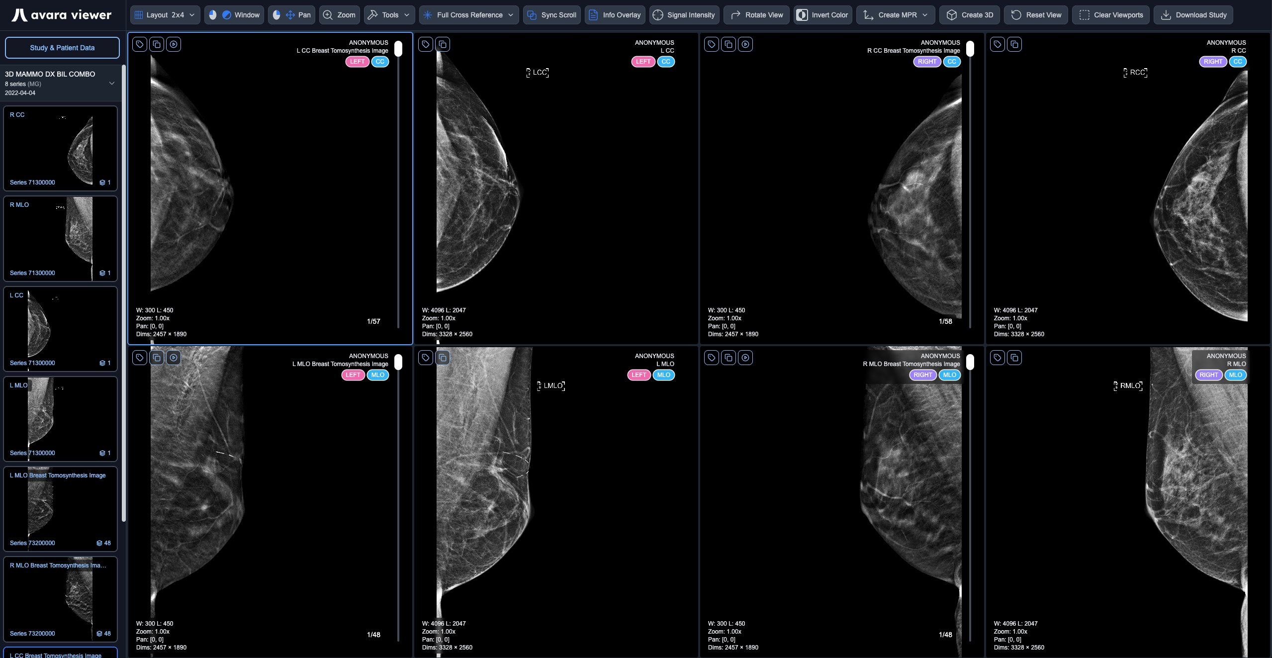
Section titled “Hanging protocol”The mammography hanging protocol uses a rules engine to determine the optimal layout and series assignment. A valid study must contain at least one series matching L CC, L MLO, R CC, or R MLO views that are not partial and do not have view modifiers (except ID, NP, or modifiers containing “synth”).
The protocol selects the most recent valid study as the current study and the next most recent valid study as the comparison study. The hanging protocol priority is checked in the following order:
- DBT current + DBT prior → 2x4 Layout: If the current study contains DBT (Digital Breast Tomosynthesis) series and a prior study also contains DBT series, use a 2x4 layout comparing current DBT to prior DBT.
- DBT current + 2D synth current (same study) → 2x4 Layout: If the current study contains both DBT and 2D synthetic series, use a 2x4 layout for internal comparison within the same study.
- DBT current + 2D non-synth current (same study) → 2x4 Layout: If the current study contains both DBT and 2D non-synthetic series, use a 2x4 layout for internal comparison within the same study.
- DBT current alone (ignore 2D priors) → 2x2 Layout: If the current study contains only DBT series with no DBT prior study, use a 2x2 layout. Any 2D prior studies are ignored.
- 2D current + 2D non-synth prior (ignore DBT priors) → 2x4 Layout: If the current study contains 2D series and a prior study contains 2D non-synthetic series, use a 2x4 layout. Any DBT prior studies are ignored.
- 2D current + 2D synth prior (ignore DBT priors) → 2x4 Layout: If the current study contains 2D series and a prior study contains 2D synthetic series, use a 2x4 layout. Any DBT prior studies are ignored.
- 2D current alone (ignore DBT priors) → 2x2 Layout: If the current study contains only 2D series with no 2D prior study, use a 2x2 layout. Any DBT prior studies are ignored.
Key constraints: DBT current studies never compare to 2D prior studies (only DBT prior or 2D within the same study), and 2D current studies never compare to DBT prior studies (only 2D prior). When multiple series match a position, the protocol prefers DBT over 2D, then synthetic vs source based on context, with lower series number as a tie-breaker.
The hanging protocol may yield between 0 and 6 empty viewports depending on the nature of the series available in the study. This is expected behavior – not a bug – as the protocol only assigns series that match the required view positions (L CC, L MLO, R CC, R MLO). If a study doesn’t contain all four standard views, the corresponding viewports will remain empty.
Display tags
Section titled “Display tags”Each mammography series displays specialized colored tags in the top right corner of the viewport, providing quick visual identification of key series attributes. The tags render in the following order:
- LATERALITY - Indicates which breast: Left (pink) or Right (purple)
- VIEW - The imaging view type: CC or MLO (blue)
- VIEW-MODIFIER - Additional view information if present (green)
- PARTIAL - Only shown if present: Partial view indicator (yellow)
These color-coded tags enable radiologists to quickly identify and distinguish between different series and views at a glance, improving workflow efficiency during mammography reading sessions.

Ex. This tag display is for a left CC implant displaced partial view
If you are wiring a PACS or platform so studies open in Avara Viewer, see Loading Viewer Data.